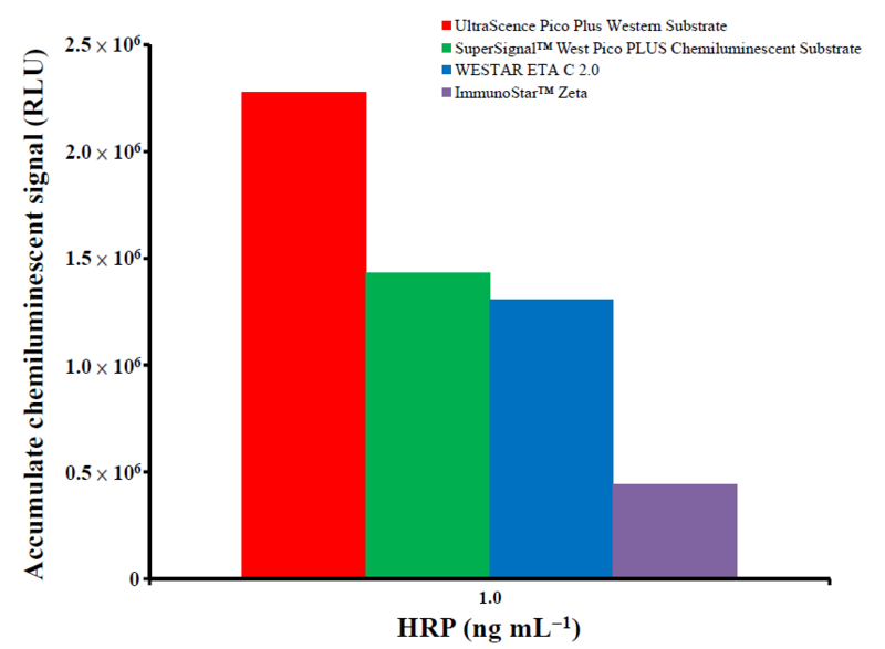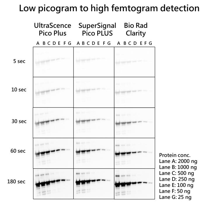
Figure 1. UltraScence Pico Plus Western Substrate enables an accurate low picogram to high femtogram detection of protein on the same immunoblot after a single exposure. Membranes were probed with GFP tag Rabbit PolyAb (Cat No. 50430-2-AP, 1:10,000) of and then with Goat Anti-rabbit IgG/HRP secondary antibody (Cat No. SA00001-2, 1:10,000) after serial dilution EGFP (Enhanced Green Fluorescent Protein) were prepared and applied in electrophoresis and protein transfer. Identical blots were incubated with the Western substrate. The blots were simultaneously exposed for 5 seconds, 10 seconds, 30 seconds, 60 seconds, and 180 seconds using ChemluxSPX-600 Series digital imaging system.
* SuperSignal West Pico PlUS and Clarity Western ECL Substrate is a registered trademark of Thermo Fisher Scientific and Bio rad. The trademark holder is not affiliated with Bio-HeliX Co., Ltd. and does not recognize this product.
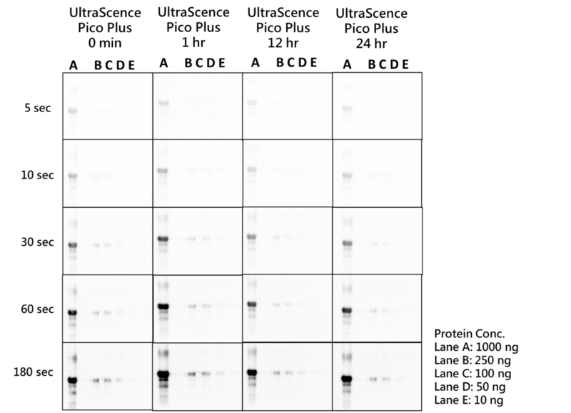
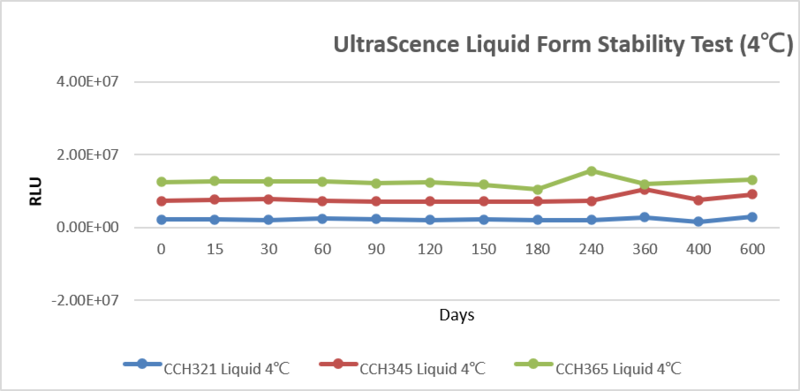
Figure 2. 24-hour stability test of UltraScence Pico Plus Western Substrate
The Luminol and Peroxide solution of UltraScence Pico Plus were mixed in a 1:1 ratio and having 400 uL treated on each blot after taken out from 4�J for 1hr, 12 hrs and 24 hrs. The blots were simultaneously exposed for 5, 10, 30, 60, and 180 seconds using Chemlux SPX-600 Series digital imaging system. The EGFP (Enhanced Green Fluorescent Protein) was serially diluted for electrophoresis, after the proteins were transferred to the membrane, they were blocked and probed with GFP tag Rabbit PolyAb (Cat No. 50430-2-AP, 1:10,000), and followed by the Goat Anti-rabbit IgG/HRP secondary antibody (Cat No. SA00001-2, 1:10,000).
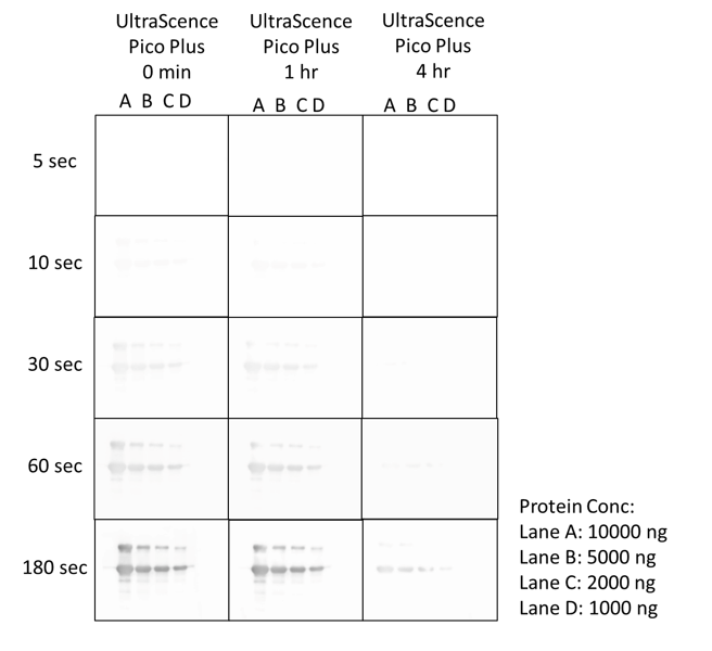
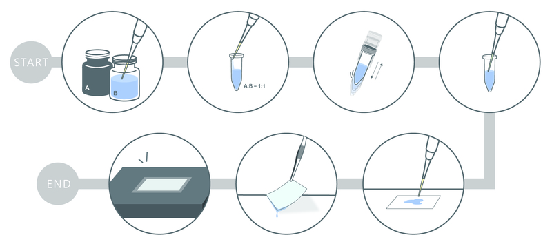
Figure 3. 4-hour signal duration with UltraScence Pico Plus Western Substrate
The EGFP (Enhanced Green Fluorescent Protein) was serially diluted for electrophoresis, after the proteins were transferred to the membrane, they were blocked and probed with GFP tag Rabbit PolyAb (Cat No. 50430-2-AP, 1:10,000), and followed by the Goat Anti-rabbit IgG/HRP secondary antibody (Cat no. SA00001-2, 1:10,000). Immerse the membrane in a mixed luminol and peroxide solution and check the signal at different times. The blots were simultaneously exposed for 5, 10, 30, 60, and 180 seconds using the Chemlux SPX-600 Series digital imaging system.